
All Images
News Release 17-069
NSF issues awards to advance a national research infrastructure for neuroscience
NeuroNex projects will develop new tools, partnerships to understand the brain
This material is available primarily for archival purposes. Telephone numbers or other contact information may be out of date; please see current contact information at media contacts.
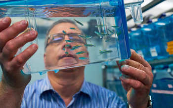
Cornell University's Joseph Fetcho will work to develop a noninvasive recording of neural activity.
Credit: Cornell University
Download the high-resolution JPG version of the image. (6.7 MB)
Use your mouse to right-click (Mac users may need to Ctrl-click) the link above and choose the option that will save the file or target to your computer.
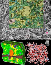
From the research of Kristen M. Harris, of the University of Texas at Austin: Electron micrograph and 3-D reconstructions from a volume equal to a single red blood cell in the hippocampus, a brain region crucial for learning and memory.
Credit: Kristen M. Harris, University of Texas at Austin
Download the high-resolution PNG version of the image. (2.1 MB)
Use your mouse to right-click (Mac users may need to Ctrl-click) the link above and choose the option that will save the file or target to your computer.
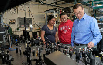
Cornell University neuroscientist Chris Xu will study how brains produce behavior in species across a range of sizes.
Credit: Cornell University
Download the high-resolution JPG version of the image. (9.8 MB)
Use your mouse to right-click (Mac users may need to Ctrl-click) the link above and choose the option that will save the file or target to your computer.
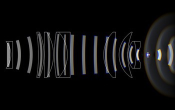
This illustration, from the lab of Spencer Smith of the University of North Carolina at Chapel Hill, shows how light propagates through an imaging objective designed for multiphoton imaging over multiple cortical areas with subcellular resolution. Light enters at the left, is focused through several lenses, and is brought to a focus just past a coverslip on the right.
Credit: Spencer L. Smith lab, slab.org
Download the high-resolution PNG version of the image. (562.9 KB)
Use your mouse to right-click (Mac users may need to Ctrl-click) the link above and choose the option that will save the file or target to your computer.
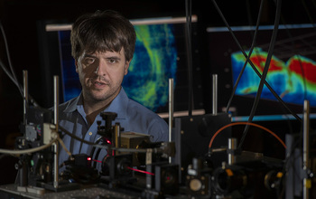
Karl Deisseroth, professor of bioengineering, psychiatry and behavioral sciences, in his lab at Stanford University.
Credit: Steve Fisch/Stanford Schoool of Medicine
Download the high-resolution JPG version of the image. (240.8 KB)
Use your mouse to right-click (Mac users may need to Ctrl-click) the link above and choose the option that will save the file or target to your computer.
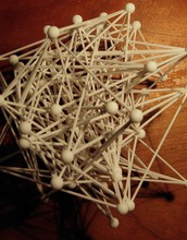
An abstract 3-D model of connected neurons, sculpted by neuroscientist Xaq Pitkow, of the Baylor College of Medicine. Pitkow is co-principal investigator for the NeuroNex award "Inferring interactions between neurons, stimuli and behavior," for which Kresimir Josic of the University of Houston is principal investigator. Identifying the connections between neurons and their functional role is the core goal of the Houston-based NeuroNex center.
Credit: Xaq Pitkow
Download the high-resolution JPG version of the image. (1.1 MB)
Use your mouse to right-click (Mac users may need to Ctrl-click) the link above and choose the option that will save the file or target to your computer.