
All Images
News Release 15-088
Bold new brain research in neuroengineering, brain-inspired design, and individuality
$13.1 million for 16 new awards part of NSF's support for integrative, fundamental brain research and the BRAIN Initiative
This material is available primarily for archival purposes. Telephone numbers or other contact information may be out of date; please see current contact information at media contacts.
Much of how brain cells connect to create meaningful networks a mystery. One NSF-funded team seeks to develop a theory of how single neural cells form electrically active networks. The project will integrate emerging methods in computer science, systems biology, neuroengineering and developmental biology to offer insight into the brain's organization.
This animation shows how young neurons develop over a two-day span.
Credit: Arun Mahadevan, Qutub Lab, Department of Bioengineering, Rice University
Download the high-resolution GIF version of the image. (5.1 MB)
Use your mouse to right-click (Mac users may need to Ctrl-click) the link above and choose the option that will save the file or target to your computer.

Brain-machine interfaces read signals directly from the brain to control external devices such as robotic limbs. While this technology has great potential to benefit people who are paralyzed, the interfaces often have poor performance because they use low-level signals to simultaneously control many aspects of the robotic limb's movements. New NSF-funded research will leverage expertise across diverse fields to generate significant improvements in brain-machine interface technology. Shown here is Erik Sorto using a brain-controlled robotic arm to take a drink.
Credit: Spencer Kellis and Christian Klaes
Download the high-resolution JPG version of the image. (81.0 KB)
Use your mouse to right-click (Mac users may need to Ctrl-click) the link above and choose the option that will save the file or target to your computer.

To capture the activity between neurons as animals move about, researchers will turn an inexpensive, lightweight tube into a super-resolution fluorescence microscope. The new tool is intended to help researchers peer deep inside the brains of freely moving mice. The new technology could help explain the fundamental basis of information processing and memory. Shown here is a genetically encoded reporter system to "tag" activated synapses, the junctions between neurons.
Credit: Andrew Taibi and Jason Shepherd, Department of Neurobiology and Anatomy, University of Utah
Download the high-resolution JPG version of the image. (52.5 KB)
Use your mouse to right-click (Mac users may need to Ctrl-click) the link above and choose the option that will save the file or target to your computer.
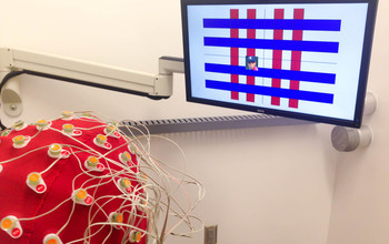
How does sleep affect memory? Researchers will monitor, characterize and manipulate memory reactivation during sleep to will expand our understanding of how memories are stored in the brain. Shown here is a person learning picture-sound associations while EEG signals are recorded.
Credit: Ken Norman, Princeton University
Download the high-resolution JPG version of the image. (486.4 KB)
Use your mouse to right-click (Mac users may need to Ctrl-click) the link above and choose the option that will save the file or target to your computer.
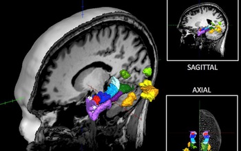
A multidisciplinary team of investigators in cognitive science, neuroscience and computer vision will examine how visual memories are encoded in the human brain at milliseconds and millimeters-resolution. The work may help predict what people remember when they see an image or an event.
Credit: Aude Oliva, MIT
Download the high-resolution JPG version of the image. (177.6 KB)
Use your mouse to right-click (Mac users may need to Ctrl-click) the link above and choose the option that will save the file or target to your computer.
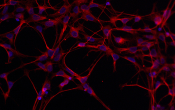
Understanding how brain cells form can tell us how the structure of neural networks relates to their function, which is an important unsolved problem in neuroengineering.
Shown here are young cells (red) two weeks into their transformation into neurons. Cell nuclei are in blue.
Credit: Arun Mahadevan, Qutub Lab, Department of Bioengineering, Rice University
Download the high-resolution JPG version of the image. (312.6 KB)
Use your mouse to right-click (Mac users may need to Ctrl-click) the link above and choose the option that will save the file or target to your computer.