
Multimedia Gallery
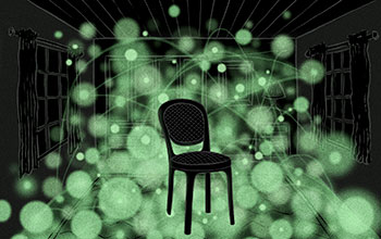
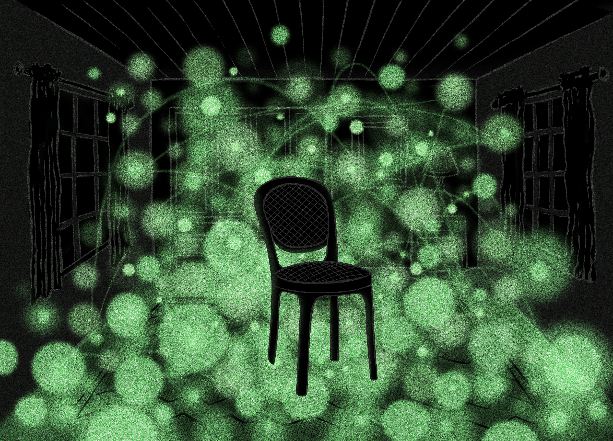
Microscope builds 3-D images by mapping negative space
Scientists at the University of Texas at Austin have developed a new microscopy technique for imaging structures in biological materials at the nanoscale that is analogous to using a glowing rubber ball to image a chair in a room in total darkness.
More about this image
Led by physicist Ernst-Ludwig Florin, a team of researchers at The University of Texas at Austin (UT-Austin) demonstrated a method called "thermal noise imaging" for making 3-D images of structures in biological material under natural conditions and at much higher resolution than other existing methods. The original concept for the thermal noise imaging technique was published and patented in 2001, but technical challenges prevented it from being developed into a fully functioning process until now. The method may shed light on how cells communicate with one another and provide important insights for engineers working to develop artificial organs such as skin or heart tissue.
The researchers used the method to capture nanometer-scale images of networks of collagen fibrils, which form part of the connective tissue found in the skin of animals. A nanometer is a billionth of a meter, or about one-hundred-thousandth of the width of a human hair. Examining collagen fibrils at this scale allowed the team to measure for the first time key properties that affect skin's elasticity, something that could lead to improved designs for artificial skin or tissues.
It is difficult to take crisp, 3-D images of nanoscale structures in biological samples partly because they tend to be soft and bathed in liquid. Tiny fluctuations in heat can cause structures to move back and forth too, an effect known as Brownian motion. To overcome the blurriness this creates, Florin and his team added nanospheres--nanometer-sized beads that reflect laser light--to their biological samples under natural conditions, shone a laser on the sample and compiled superfast snapshots of the nanospheres viewed through a light microscope. This is the thermal noise imaging method.
The thermal noise imaging method works something like the analogy pictured in this image: Imagine you needed to take a 3-D image of a room in total darkness. If you were to throw a glowing rubber ball into the room and use a camera to collect a series of high-speed images of the ball as it bounces around, you would see that as the ball moves around the room, it isn't able to move through solid objects such as tables and chairs. Combining millions of images taken so fast that they don't blur, you would be able to build a picture of where there are objects (wherever the ball couldn't go) and where there aren't objects (where it could go). In thermal noise imaging, the equivalent of the rubber ball is a nanosphere that moves around in a sample by natural Brownian motion.
"If you want to build artificial skin, you have to understand how the natural components work," says Florin. "You could then better design a collagen network that acts as a scaffolding that encourages cells to grow in the right way."
This research was supported in part by National Science Foundation grants PHY 06-47144, DBI 05-52094 and DMR 14-11262.
To learn more about this research, see the UT-Austin news story Super-resolution Microscope Builds 3-D Images by Mapping Negative Space. (Date image taken: September 2016; date originally posted to NSF Multimedia Gallery: Feb. 14, 2017)
Credit: Jenna Luecke, The University of Texas at Austin
See other images like this on your iPhone or iPad download NSF Science Zone on the Apple App Store.
Images and other media in the National Science Foundation Multimedia Gallery are available for use in print and electronic material by NSF employees, members of the media, university staff, teachers and the general public. All media in the gallery are intended for personal, educational and nonprofit/non-commercial use only.
Images credited to the National Science Foundation, a federal agency, are in the public domain. The images were created by employees of the United States Government as part of their official duties or prepared by contractors as "works for hire" for NSF. You may freely use NSF-credited images and, at your discretion, credit NSF with a "Courtesy: National Science Foundation" notation.
Additional information about general usage can be found in Conditions.
Also Available:
Download the high-resolution JPG version of the image. (5.2 MB)
Use your mouse to right-click (Mac users may need to Ctrl-click) the link above and choose the option that will save the file or target to your computer.