
Multimedia Gallery
 All images in this series
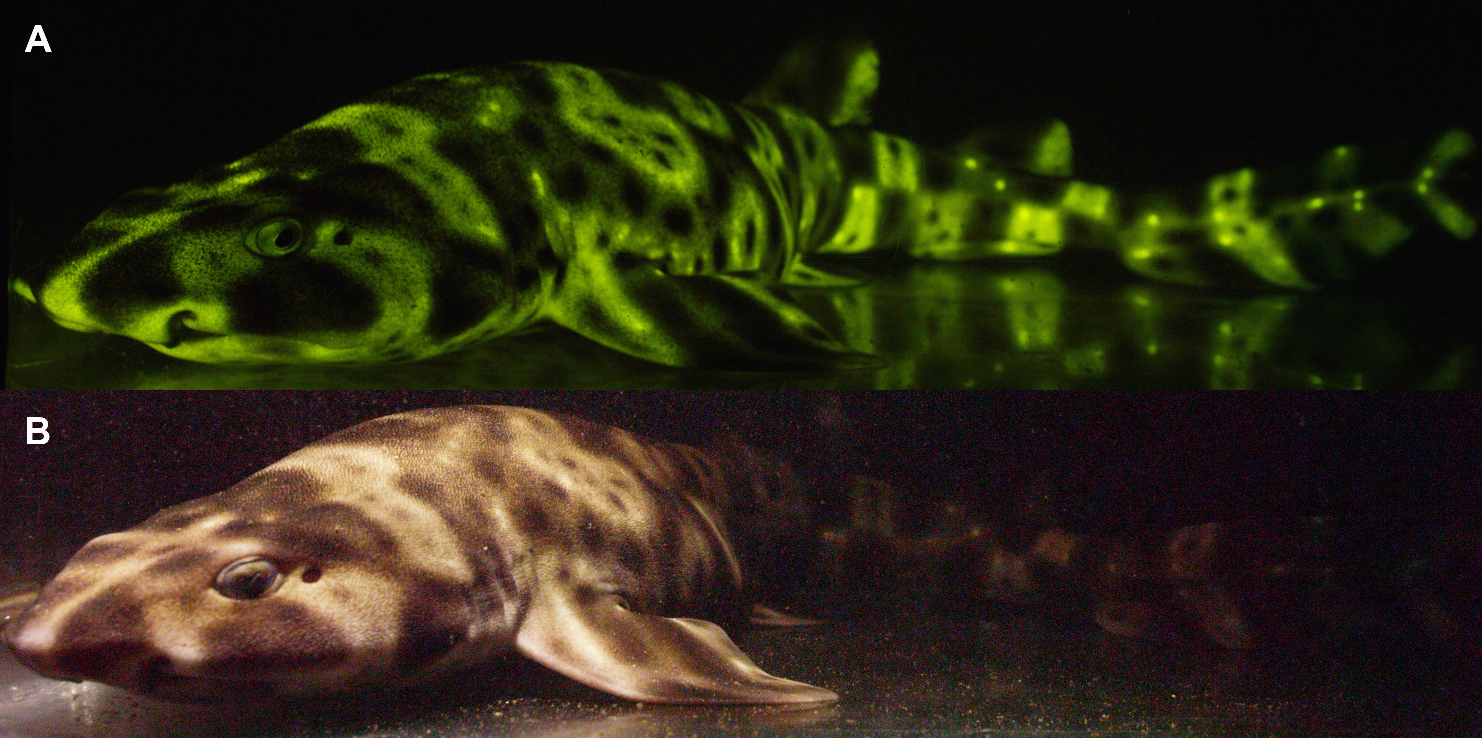
A fluorescent and white light image of a female swellshark
A fluorescent (A) and white light (B) image of a female swellshark (Cephaloscyllium ventriosum).
More about this image
A study led by researchers at the American Museum of Natural History has found that catsharks (Scyliorhinus rotifer) not only see the bright green biofluorescence they produce, but they can increase contrast of their glowing pattern when deep underwater.
The researchers found that fluorescence makes catsharks more visible to neighbors of the same species at the depths that they live and may aid in communication between them.
A paper published about the study says it "is one of the first papers on biofluorescence to show a connection between visual capability and fluorescence emission, and a big step toward a functional explanation for fluorescence in fishes," said John Sparks, a curator in AMNH's Department of Ichthyology and co-author of the paper. Biofluorescence is a phenomenon in which organisms absorb light, transform it and emit it as a different color.
Fish live in a mostly "blue" world. As you go deeper in the ocean, water quickly absorbs the majority of the visible light spectrum. Previous studies by the team found that many fish absorb the remaining blue light and re-emit it in neon greens, reds and oranges. To capture this hidden biofluorescent world, the researchers designed lighting that mimics the ocean’s light, along with cameras that can capture the animals’ fluorescent light. (The researchers also recently made the first observation of biofluorescence in marine turtles.)
The researchers sought to find out what this biofluorescence in the ocean means and, more specifically, can the animals see other animals that are biofluorescing in the deep ocean and are they using it in some manner?
To explore the phenomenon, the researchers focused on the visual ability of two catsharks: chain catsharks (Scyliorhinus rotifer) and swellsharks (Cephaloscyllium ventriosum). Working in collaboration with Cornell University veterinary expert Ellis Loew, the researchers used a technique called microspectrophotometry to determine how the sharks’ eyes absorb light. They discovered the sharks have long rod pigments that help them see in low-light environments. Using this knowledge, they built a special camera filter that simulates how light hits a shark’s eyes -- a "shark-eye" camera.
For the study, David Gruber, an associate professor of biology at Baruch College and a research associate at AMNH, and Sparks observed swellsharks in their native habitat, about 100 feet under water at Scripps Canyon in San Diego County. Diving at night, the team used high-intensity, blue light arrays housed in watertight cases to stimulate biofluorescense in the sharks -- the resulting light show is invisible to the human eye. The researchers recorded the activity using custom-built underwater cameras with green filters (to block out the blue light), and using the newly developed "shark-eye" camera, to better understand how the sharks see the underwater display.
Next, they mathematically modeled images from the shark-eye camera and found that the contrast of patterns on the biofluorescent sharks increases with depth, suggesting that the animals can not only see the light, but are also likely using it to communicate with one another. The researchers were only able to dive to the top depth range of where this shark lives, where blue and some green light exists. Their model shows that at deeper depths, where the water is bluer, the contrast created by the fluorescence is even greater.
This research was supported in part by the U.S. National Science Foundation (grants DEB 1257555, DEB 1258141 and DBI 1040321).
To read more about this research, see the AMNH news story Patterns of glowing sharks get clearer with depth. (Date image taken: April 2016; date originally posted to NSF Multimedia Gallery: Feb. 3, 2017)
Credit: ©Gruber et al
Images and other media in the National Science Foundation Multimedia Gallery are available for use in print and electronic material by NSF employees, members of the media, university staff, teachers and the general public. All media in the gallery are intended for personal, educational and nonprofit/non-commercial use only.
Images credited to the National Science Foundation, a federal agency, are in the public domain. The images were created by employees of the United States Government as part of their official duties or prepared by contractors as "works for hire" for NSF. You may freely use NSF-credited images and, at your discretion, credit NSF with a "Courtesy: National Science Foundation" notation.
Additional information about general usage can be found in Conditions.
Also Available:
Download the high-resolution JPG version of the image. (4.1 MB)
Use your mouse to right-click (Mac users may need to Ctrl-click) the link above and choose the option that will save the file or target to your computer.