
Multimedia Gallery
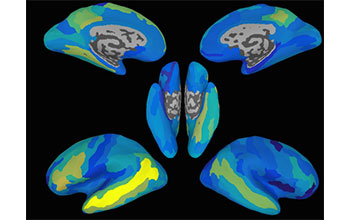
Brain maps predict neural activation patterns
These brain maps show how accurately it was possible for researchers at the University of Rochester to predict neural activation patterns for new, previously unseen sentences in different regions of the brain. The brighter the area, the higher the accuracy. The most accurate area, which can be seen as the bright yellow strip, is a region in the left side of the brain known as the Superior Temporal Sulcus. This region achieved statistically significant sentence predictions in 11 out of the 14 people whose brains were scanned. Although that was the most accurate region, several other regions, broadly distributed across the brain, also produced significantly accurate sentence predictions.
More about this image
Researchers at the University of Rochester have for the first time decoded and predicted the brain activity patterns of word meanings within sentences, and successfully predicted what the brain patterns would be for new sentences.
The study used functional magnetic resonance imaging (fMRI) to measure human brain activation. "Using fMRI data, we wanted to know if given a whole sentence, can we filter out what the brain’s representation of a word is--that is to say, can we break the sentence apart into its word components, then take the components and predict what they would look like in a new sentence," said Andrew Anderson, a research fellow who led the study as a member of the lab of Rajeev Raizada, assistant professor of brain and cognitive sciences at Rochester.
"We found that we can predict brain activity patterns--not perfectly [on average 70 percent correct], but significantly better than chance," said Anderson. Anderson and his colleagues say the study makes key advances toward understanding how information is represented throughout the brain.
The research was supported in part by the National Science Foundation (grant BCS 12-28261).
To learn more, see the NSF News From the Field story This is your brain on sentences (Date image taken: 2016; date originally posted to NSF Multimedia Gallery: Jan. 25, 2017)
Credit: University of Rochester graphic / Andrew Anderson and Xixi Wang
Images and other media in the National Science Foundation Multimedia Gallery are available for use in print and electronic material by NSF employees, members of the media, university staff, teachers and the general public. All media in the gallery are intended for personal, educational and nonprofit/non-commercial use only.
Images credited to the National Science Foundation, a federal agency, are in the public domain. The images were created by employees of the United States Government as part of their official duties or prepared by contractors as "works for hire" for NSF. You may freely use NSF-credited images and, at your discretion, credit NSF with a "Courtesy: National Science Foundation" notation.
Additional information about general usage can be found in Conditions.
Also Available:
Download the high-resolution JPG version of the image. (251.1 KB)
Use your mouse to right-click (Mac users may need to Ctrl-click) the link above and choose the option that will save the file or target to your computer.