
Multimedia Gallery
 All images in this series
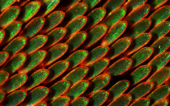
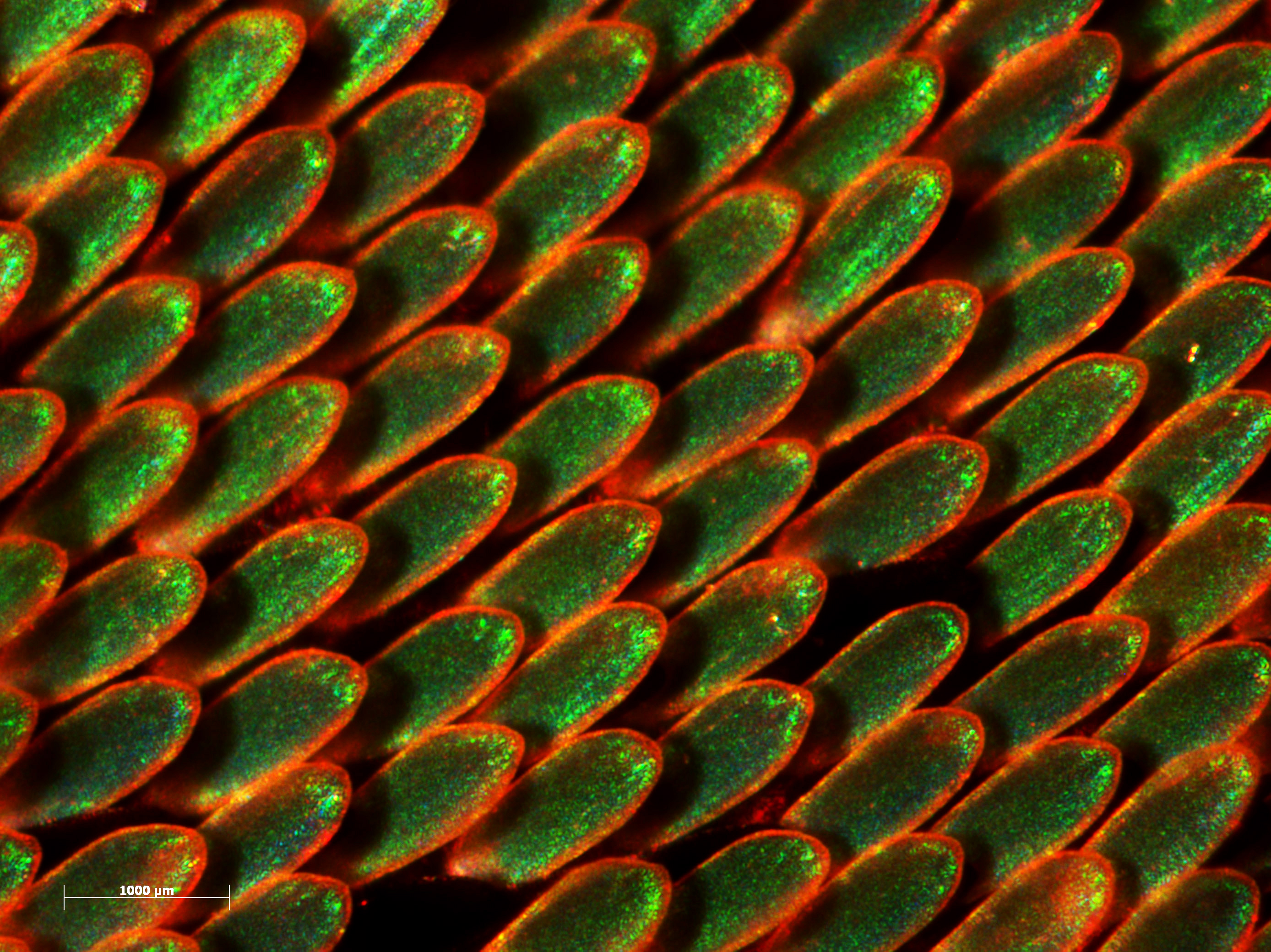
Emerald green scales on wings of emerald-patched cattleheart butterfly
Like shingles on a roof, vivid emerald-green scales adorn the wings of the Amazonian butterfly emerald-patched cattleheart.
More about this Image
Researchers from Yale University, supported by the National Science Foundation, are studying the properties of the colors of butterfly wings. Using an X-ray scattering technique, they were able to determine the 3-D internal structure of the scales on the wings of several species of butterflies.
The crystal nanostructures that give butterflies their color are called gyroids--strange, 3-D curving structures that selectively scatter light. The gyroids are made of chitin (the same tough, starchy material that forms the exoskeletons of insects and crustaceans), which is usually deposited on the outer membranes of cells. Researchers wanted to know how the cells sculpt themselves into these extraordinary forms resembling a network of three-bladed boomerangs. They discovered that the outer membranes of the butterfly wing scale cells grow and fold into the interior of the cells. The membranes then form a double gyroid--two, mirror-image networks shaped by the outer and inner cell membranes. The latter are easier to grow but are not as good at scattering light. The chitin is then deposited in the outer gyroid to create a single solid crystal. After this the cell dies, leaving behind the crystal nanostructures on the butterfly wing.
Photonic engineers are using gyroid shapes to try and create more efficient solar cells and, by mimicking nature, may be able to produce more efficient optical devices as well.
[This butterfly research was supported in part by a seed grant from Yale University's Center for Research on Interface Structures and Phenomena (CRISP) (under National Science Foundation grant DMR 05-20495)--an NSF Materials Research Science and Engineering Center (MRSEC), and by NSF grants to Simon G.J. Mochrie, department of physics, and Richard O. Prum, department of ornithology, ecology and evolutionary biology, Yale University.] (Date of Image: August 2009)
Credit: Richard Prum, Department of Ecology and Evolutionary Biology and Peabody Museum of Natural History, Yale University
See other images like this on your iPhone or iPad download NSF Science Zone on the Apple App Store.
Images and other media in the National Science Foundation Multimedia Gallery are available for use in print and electronic material by NSF employees, members of the media, university staff, teachers and the general public. All media in the gallery are intended for personal, educational and nonprofit/non-commercial use only.
Images credited to the National Science Foundation, a federal agency, are in the public domain. The images were created by employees of the United States Government as part of their official duties or prepared by contractors as "works for hire" for NSF. You may freely use NSF-credited images and, at your discretion, credit NSF with a "Courtesy: National Science Foundation" notation.
Additional information about general usage can be found in Conditions.
Also Available:
Download the high-resolution JPG version of the image. (3.0 MB)
Use your mouse to right-click (Mac users may need to Ctrl-click) the link above and choose the option that will save the file or target to your computer.