
Multimedia Gallery
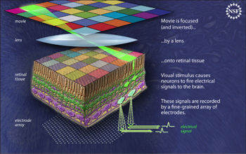
Updated retinal readout system can record signals from hundreds of cells simultaneously.
To understand how individual retinal ganglion cells in the eye work together to send signals that the brain interprets as color vision, scientists studied the pattern of connections and signals that occur when a movie is projected on live retinal tissue. The tissue sits on top of a multi-electrode array that detects the signals the cells send to the brain.
In contrast to earlier technology, which utilized a small number of electrodes that could only monitor signals from tens of cells, the retinal readout system allows simultaneous recording of signals from hundreds of cells. These signals are filtered, digitized and recorded, then related to the visual images in the movie. This advanced technology is helping the researchers decipher the code that eyes use to send visual information to the brain.
Credit: Zina Deretsky, National Science Foundation
Images credited to the National Science Foundation, a federal agency, are in the public domain. The images were created by employees of the United States Government as part of their official duties or prepared by contractors as "works for hire" for NSF. You may freely use NSF-credited images and, at your discretion, credit NSF with a "Courtesy: National Science Foundation" notation.
Additional information about general usage can be found in Conditions.
Also Available:
Download the high-resolution JPG version of the image. (551 KB)
Use your mouse to right-click (Mac users may need to Ctrl-click) the link above and choose the option that will save the file or target to your computer.
Related story: Mapping Color Vision in HD