
Multimedia Gallery
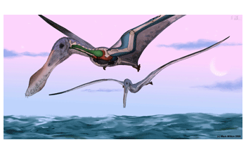
Pterosaur Anhanguera
An artist's conception showing the distribution of balloon-like air sacs of the four-meter wingspan pterosaur, Anhanguera. The air sacs extended from the lungs to inside the skeleton of pterosaurs, providing an efficient breathing system for these ancient beasts. [Colors denote differentiated parts of the respiratory system: lungs (orange), cervical air sacs (green), abdominal air sacs (blue), thoracic air sacs (gray) and subcutaneous diverticular network extending along the leading edge of the wing (light blue).]
More about this Image
This information was published in a study by researchers from Ohio University, the College of the Holy Cross and the University of Leicester. Leon Claessens, assistant professor of biology at the College of the Holy Cross, and Patrick O'Connor, assistant professor of biomedical sciences at the Ohio University College of Osteopathic Medicine, came up with the idea for the study after meeting with David Unwin of the University of Leicester, who was curator at the Natural History Museum in Berlin at the time. Unwin showed them an extraordinarily preserved pterosaur, which the scientists thought that by studying, might finally explain how the pterosaur powered sustained flight.
"The shape and size of the rib segments that articulate with the sternum indicate that the ribcage was mobile, contrary to previous ideas," Claessens said. He added that the unique and previously unrecognized projections on the ribs provided important leverage for the muscles that power lung ventilation.
Fossils rarely preserve soft tissues, so the research team conducted a comparative study that included pterosaurs, birds and crocodilians, to gain a better understanding of the relationships among air sacs, lung structure and the skeleton. The researchers used X-ray movies and CT scans in order to characterize how the skeleton works to move air through the lungs in living animals, and also how to identify the signature traces left on bones that have been invaded by air sacs.
In addition to extinct pterosaurs showing evidence that their bones were invaded by air sacs, there are patterns of pneumaticity throughout the entire skeleton of different pterosaur species that parallel trends identified in many living bird groups. For example, there is a direct relationship between the proportion of the skeleton invaded by air sacs and the absolute body size of an animal.
"Whereas small-bodied pterosaurs and birds typically pneumatize only a restricted part of the backbone, larger-bodied species routinely pegmatite most bones of the body, including the wing skeleton out to the ends of the fingers," O'Connor said. Such modifications of the skeleton would have reduced bone density and resolved a major problem with sustaining flight in large-bodied pterosaurs: the energetic cost of keeping a heavy body up in the air. Density reduction of the skeleton in pterosaurs may have been beneficial, particularly so in these aerial giants, just as it appears to be in the largest flying birds today. The existence of an analogous air-sac system in pterosaurs highlights new areas of research in which paleobiologists can explore aspects of pterosaurian biology. [Research funded by the National Science Foundation, Harvard University and the Ohio University College of Osteopathic Medicine and Office of Research. Story information taken from Ohio University news release, "Air-filled bones helped prehistoric reptiles take first flight."] (Date of Image: November 2009)
Credit: Mark Witton
See other images like this on your iPhone or iPad download NSF Science Zone on the Apple App Store.
Special Restrictions: This work by Mark Witton is licensed under a Creative Commons (CC) Attribution-Noncommercial-Share Alike 2.0 Generic Creative Commons (CC) License, whereby you are allowed to (1) copy, distribute, display and perform the work; or (2) make derivative works, under the condition that (a) you give the original author credit; (b) you may not use this work for commercial purposes; and (c) if you alter, transform or build upon this work, you may distribute the resulting work only under a license identical to this one. For more information, visit the CC website Here.
Images and other media in the National Science Foundation Multimedia Gallery are available for use in print and electronic material by NSF employees, members of the media, university staff, teachers and the general public. All media in the gallery are intended for personal, educational and nonprofit/non-commercial use only.
Images credited to the National Science Foundation, a federal agency, are in the public domain. The images were created by employees of the United States Government as part of their official duties or prepared by contractors as "works for hire" for NSF. You may freely use NSF-credited images and, at your discretion, credit NSF with a "Courtesy: National Science Foundation" notation.
Additional information about general usage can be found in Conditions.
Also Available:
Download the high-resolution JPG version of the image. (3.4 MB)
Use your mouse to right-click (Mac users may need to Ctrl-click) the link above and choose the option that will save the file or target to your computer.