
Multimedia Gallery

Human Face Recognition Study (Image 4)
Human Face Recognition Study (Image 4)
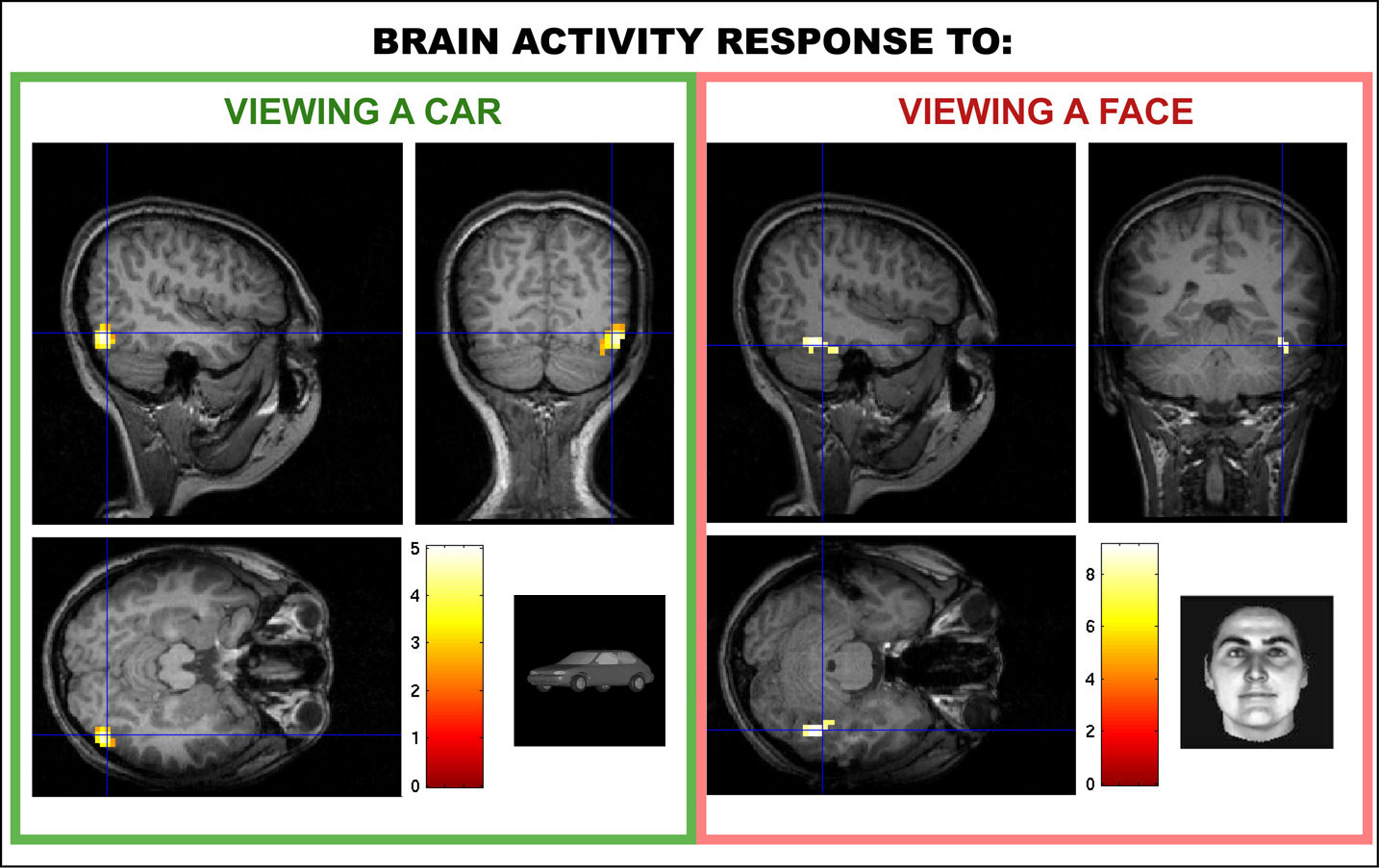
Example of an ongoing fMRI study investigating how learning an object recognition task (in this case, categorization of cars generated with a computer graphics morphing system) 'reprograms' the brain to improve performance on the task. The left part of the figure shows a brain area found in trained subjects that is selective to cars, very much in the same way the "fusiform face area" shown in the right is selective for human faces. Image courtesy of NSF based on our data. [Note: The color scale shows a measure of statistical significance (z-value). The lighter the color, the more significant the activation. The blue lines are drawn for illustration to show the center of the region of interest.]
This research was conducted by Maximilian Riesenhuber, a neuroscientist at Georgetown University Medical Center, who is working to better understand how the human brain functions when recognizing objects. He published data in the April 6 issue of Neuron that suggested the human brain uses the same mechanisms for recognizing face and non-face objects.
Riesenhuber's research is partly funded by the Collaborative Research in Computational Neuroscience (CRCNS) program, a joint effort of the National Science Foundation and the National Institutes of Health. The program supports innovative, interdisciplinary research that will yield a better understanding of how nervous systems function normally as well as when diseased. By fostering new collaborations among computer scientists, engineers, mathematicians and neuroscientists, CRCNS facilitates the development of new methods and computational tools to help explain complex biological processes.
For more information on Riesenhuber's facial recognition research, visit his Web site, The Laboratory for Computational Cognitive Neuroscience. (Date of Image: March 2006) [Image 4 of 4 related images. Back to Image 1.]
Credit: Maximilian Riesenhuber, Georgetown University Medical Center
Images and other media in the National Science Foundation Multimedia Gallery are available for use in print and electronic material by NSF employees, members of the media, university staff, teachers and the general public. All media in the gallery are intended for personal, educational and nonprofit/non-commercial use only.
Images credited to the National Science Foundation, a federal agency, are in the public domain. The images were created by employees of the United States Government as part of their official duties or prepared by contractors as "works for hire" for NSF. You may freely use NSF-credited images and, at your discretion, credit NSF with a "Courtesy: National Science Foundation" notation.
Additional information about general usage can be found in Conditions.
Also Available:
Download the high-resolution JPG version of the image. (451 KB)
Use your mouse to right-click (Mac users may need to Ctrl-click) the link above and choose the option that will save the file or target to your computer.