
Multimedia Gallery
"Light Scattered by Gold Nanorods"
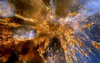
"Light Scattered by Gold Nanorods."
In this image, gold nanorods, embedded in a cell-populated collagen gel, scatter light as viewed under a darkfield microscope. The collective excitation of electrons in the conduction band of gold nanoparticles arising from resonance with incident-visible radiation is referred to as localized surface plasmon resonance. This excitation leads to resonant Rayleigh light scattering. Because of this strong scattering, individual nanoparticles, much smaller than the wavelength of light, can be observed using an optical microscope. There has been considerable interest in resonant Rayleigh scattering from gold and silver nanoparticles for biological and chemical analysis. In this application, a fibroblast-seeded collagen gel, an in vitro material system often used to model wound healing, is embedded with nanoparticles. The pattern of scattered light will be tracked, using computerized pattern matching and image correlation techniques, to measure the deformation that occurs as the collagen gel contracts, in a simulation of the formation of scar tissue. It is hoped that these small scale measurements will illustrate local heterogeneity in the mechanical response of the material.
This image is part of a proof-of-concept experiment for an interdisciplinary project between engineers, chemists, cell biologists and artists from three schools and the University of South Carolina, the College of Arts and Sciences, the College of Engineering and Information Technology and the School of Medicine. It was an entry in the 2005 Science & Engineering Visualization Challenge competition, sponsored by the National Science Foundation and the journal Science. The competition is held each year to recognize outstanding achievements by scientists, engineers, visualization specialists and artists who are innovators in using visual media to promote the understanding of research results and scientific phenomena. To learn more about the competition and view all the winning entries, see the Science & Engineering Visualization Challenge Special Report. (Date of Image: February 2004)
Credit: The USC Nanocenter
See other images like this on your iPhone or iPad download NSF Science Zone on the Apple App Store.
Images and other media in the National Science Foundation Multimedia Gallery are available for use in print and electronic material by NSF employees, members of the media, university staff, teachers and the general public. All media in the gallery are intended for personal, educational and nonprofit/non-commercial use only.
Images credited to the National Science Foundation, a federal agency, are in the public domain. The images were created by employees of the United States Government as part of their official duties or prepared by contractors as "works for hire" for NSF. You may freely use NSF-credited images and, at your discretion, credit NSF with a "Courtesy: National Science Foundation" notation.
Additional information about general usage can be found in Conditions.
Also Available:
Download the high-resolution TIF version of the image. (18.8 MB)
Use your mouse to right-click (Mac users may need to Ctrl-click) the link above and choose the option that will save the file or target to your computer.