
Multimedia Gallery

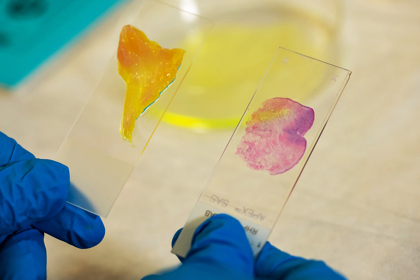
Large tissue sections imaged using microscope with AI technology
The deep learning extended depth-of-field microscope uses artificial intelligence to quickly image all the cells in large tissue sections (left) at high resolution and with minimal preparation, eliminating the process of mounting thin tissue slices on slides (right).
[Research supported by U.S. National Science Foundation grants IIS 1730574 and EEC 1648451.]
Learn more about this research in the Rice University news story AI-powered microscope could check cancer margins in minutes. (Date of image: unknown; date originally posted to NSF Multimedia Gallery: April 26, 2021)
Credit: Brandon Martin/Rice University
Images and other media in the National Science Foundation Multimedia Gallery are available for use in print and electronic material by NSF employees, members of the media, university staff, teachers and the general public. All media in the gallery are intended for personal, educational and nonprofit/non-commercial use only.
Images credited to the National Science Foundation, a federal agency, are in the public domain. The images were created by employees of the United States Government as part of their official duties or prepared by contractors as "works for hire" for NSF. You may freely use NSF-credited images and, at your discretion, credit NSF with a "Courtesy: National Science Foundation" notation.
Additional information about general usage can be found in Conditions.
Also Available:
Download the high-resolution JPG version of the image. (333.6 KB)
Use your mouse to right-click (Mac users may need to Ctrl-click) the link above and choose the option that will save the file or target to your computer.