
Multimedia Gallery
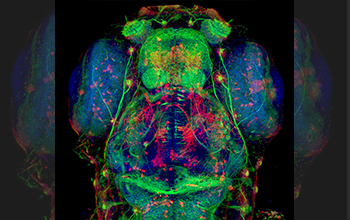
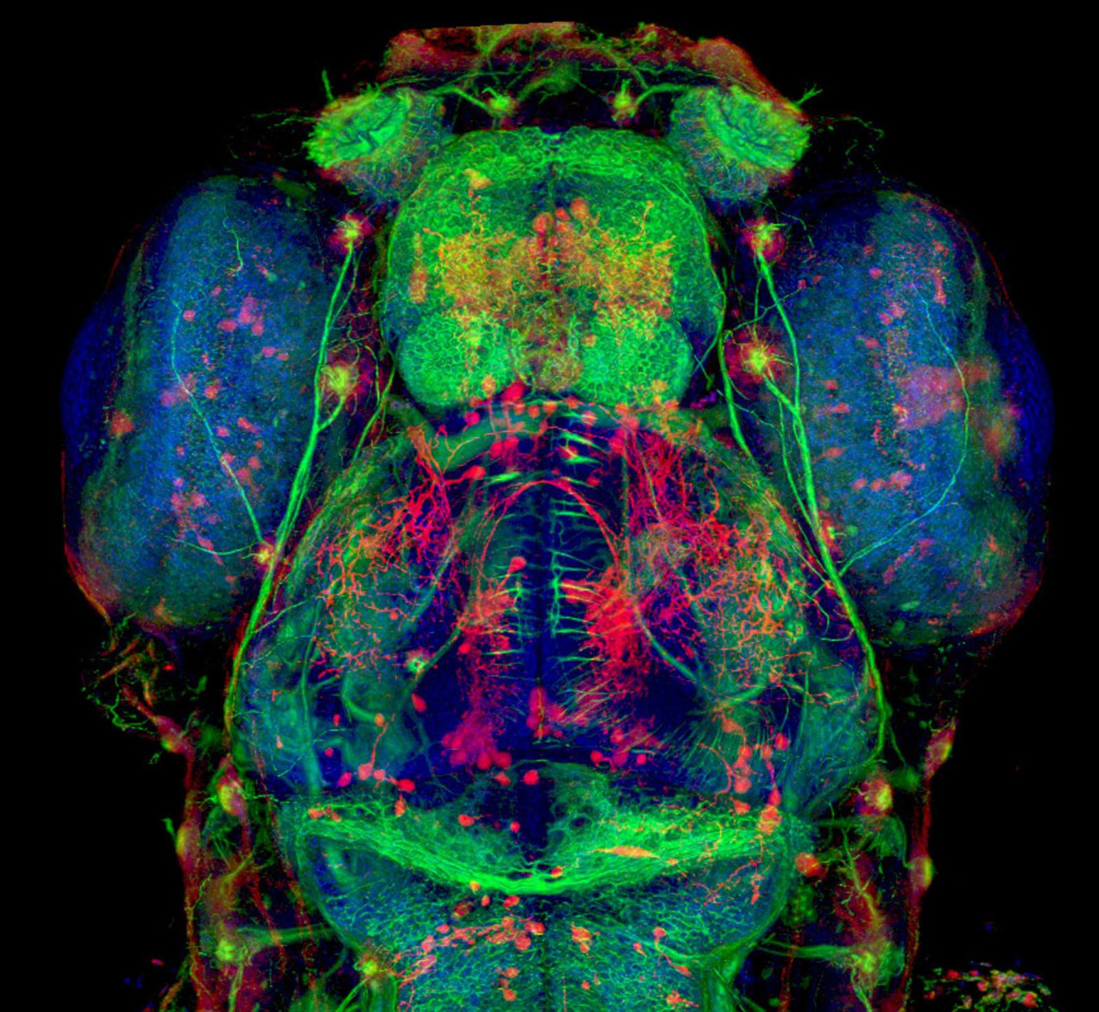
Confocal microscopy image of nerve fibers in zebrafish brain
This confocal microscopy image shows glowing red, green and blue nerve fibers in the brain of a five-day-old zebrafish.
[Research supported by National Science Foundation grants CCF 0541113, CNS 0615194, CNS 0551724, IIS 0513212 and OCI 0906379.]
This image appeared in the NSF gallery "Imaging the Imperceptible," published in 2018 in Discover magazine. (Date image taken: March 2010; date originally posted to NSF Multimedia Gallery: Dec. 17, 2019)
Credit: HHMI Janelia Research Campus
Images and other media in the National Science Foundation Multimedia Gallery are available for use in print and electronic material by NSF employees, members of the media, university staff, teachers and the general public. All media in the gallery are intended for personal, educational and nonprofit/non-commercial use only.
Images credited to the National Science Foundation, a federal agency, are in the public domain. The images were created by employees of the United States Government as part of their official duties or prepared by contractors as "works for hire" for NSF. You may freely use NSF-credited images and, at your discretion, credit NSF with a "Courtesy: National Science Foundation" notation.
Additional information about general usage can be found in Conditions.
Also Available:
Download the high-resolution JPG version of the image. (1.3 MB)
Use your mouse to right-click (Mac users may need to Ctrl-click) the link above and choose the option that will save the file or target to your computer.