
Multimedia Gallery
Tubes From Pollen Headed for an Egg Cell
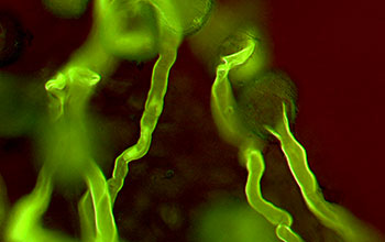
Pollen tubes emerging from pollen grains on the stylar surface of Austrobaileya scandens, a vine endemic to the Australian rainforest. The structures have been stained with aniline blue in order to visualize callose, a type of carbohydrate made up of sugar molecules that can be rapidly synthesized and secreted in plant cell walls. Callose stains bright yellow-green under ultraviolet light. The colors are created by the mix of fluorescence with faint background bright light to highlight the structural features of the pollen wall and stigma (the part of the flower that receives pollen).
A Zeiss Axioplan II light microscope equipped for fluorescence was used to both fluoresce and backlight the image at the same time. The pollen tubes exit the pollen grain from a slit-like opening that can be most clearly seen in the pollen grain on the right side. The tubes are about 10 microns wide (one-one hundredth of a millimeter), but they can grow well over 6 millimeters to reach an egg, all in less than 24 hours after pollination, which is when this photo was taken.
This research was performed by Joseph Williams, an evolutionary biologist at the University of Tennessee who is investigating why flowering plants have diversified so much more quickly than cone-bearing plants. There are an estimated 270,000 known species of angiosperms (flowering plants) but only about 900 species of gymnosperms (nonflowering plants like conifers, cycads and ginkgoes).
Williams is studying the nature of this process of getting sperm to the egg in a group of early-diverging lineages of flowering plants in order to find out if there are commonalities in the fertilization process among them. Such commonalities would suggest ancient features of the fertilization process that cannot be studied in fossil lineages. To learn more about Williams' research, visit his website Here. [Research supported by National Science Foundation grant DEB 06-40792.] (Date of Image: August 2007)
SORRY: THIS IMAGE IS NOT AVAILABLE IN HIGH RESOLUTION FORMAT
Credit: Joseph H. Williams, Department of Ecology and Evolution, University of Tennessee
Images and other media in the National Science Foundation Multimedia Gallery are available for use in print and electronic material by NSF employees, members of the media, university staff, teachers and the general public. All media in the gallery are intended for personal, educational and nonprofit/non-commercial use only.
Images credited to the National Science Foundation, a federal agency, are in the public domain. The images were created by employees of the United States Government as part of their official duties or prepared by contractors as "works for hire" for NSF. You may freely use NSF-credited images and, at your discretion, credit NSF with a "Courtesy: National Science Foundation" notation.
Additional information about general usage can be found in Conditions.
Also Available:
Download the high-resolution TIF version of the image. (1.6 MB)
Use your mouse to right-click (Mac users may need to Ctrl-click) the link above and choose the option that will save the file or target to your computer.