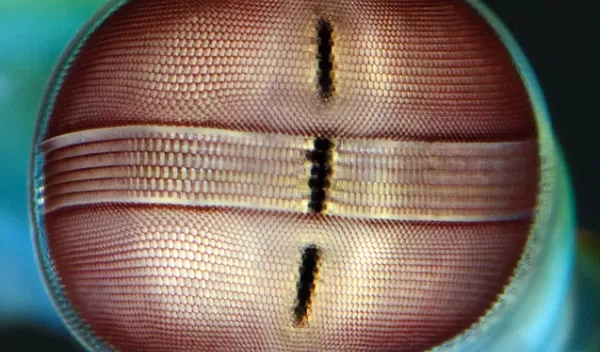
How mantis shrimp make sense of the world
A new, NSF-supported study published in the Journal of Comparative Neurology provides insight into how the small brains of mantis shrimp -- fierce predators with keen vision -- may process and integrate visual information with other sensory input.
A team of researchers examined the neuronal organization of mantis shrimp and discovered a region of their brain called the reniform body.
Nicholas Strausfeld of the University of Arizona teamed up with other researchers to better understand how the reniform bodies connect to other parts of the mantis shrimp brain.
Mantis shrimp eyes have stereoscopic vision and possess a band of photoreceptors that can distinguish up to 12 different wavelengths and linear and circular polarized light. Mantis shrimp seem to be able to process all the different channels of information with the participation of the reniform body.
A crucial finding was that neural connections link the reniform bodies to centers called mushroom bodies, iconic structures of arthropod brains that are required for olfactory learning and memory.
"The fact that we were now able to demonstrate that the reniform body is also connected to the mushroom body and provides information to it suggests that olfactory processing may take place in the context of already established visual memories," said Strausfeld.
"This work by Professor Strausfeld and his colleagues is part of a larger innovative project to better understand the organization and evolution of the brains of invertebrates. It provides a wonderful example of the unanticipated discoveries that result from applying state-of-the art research techniques to understudied organisms," said Maria Sutton of NSF's Directorate for Biological Sciences.


