
All Images
Research News
Like cotton candy? You'll love electrospinning
Researchers funded by the National Science Foundation are creating a new biosensor that uses laser light, engineered viruses and advanced manufacturing techniques to more accurately detect the smallest amounts possible of biological molecules--in our food, in our water and even in our own blood.
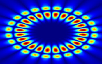
Shown here is a cross-sectional view of a circular optical cavity or resonator showing whispering gallery modes total internally reflected along the surface of a fiber.
Credit: Joe Cheeney, University of California-Riverside
Download the high-resolution JPG version of the image. (81.8 KB)
Use your mouse to right-click (Mac users may need to Ctrl-click) the link above and choose the option that will save the file or target to your computer.
One technique, known as electrospinning, creates long, hair-like fibers made of plastic, metal or ceramics. This fiber-making process is like making cotton candy, says Engineer Nosang Myung at the University of California, Riverside, who has worked with nano-sized, bio-manufactured structures for more than a decade. "You have a drop of liquid. Spin it. Out comes a long fiber. It's just like a cotton candy machine, except you apply electrical fields to spin it up," he says.
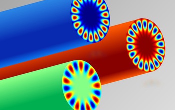
The light or electromagnetic field profile of whispering gallery modes are shown propagating along the periphery of three fiber resonators.
Credit: Joe Cheeney, University of California-Riverside
Download the high-resolution JPG version of the image. (81.5 KB)
Use your mouse to right-click (Mac users may need to Ctrl-click) the link above and choose the option that will save the file or target to your computer.
Using viruses is another new approach for biosensor technology. Not only are few--if any--biosensors created by electrospinning, most use enzymes. But enzymes are fragile and don't last long at room temperature, according to Engineers Elaine Haberer at the University of California, Riverside. Viruses have more staying power.
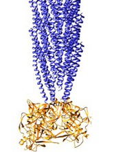
Shown here is a schematic of the filamentous M13 bacterophage or virus, which is used to organize the biorecognition elements of the biosensor.
Credit: Steven Garcia, University of California-Riverside
Download the high-resolution JPG version of the image. (112.1 KB)
Use your mouse to right-click (Mac users may need to Ctrl-click) the link above and choose the option that will save the file or target to your computer.

Engineers Elaine Haberer and Nosang Myung at the University of California, Riverside, use laser light to amplify the detection of single particles, a technique known as whispering gallery mode resonators. Whispering galleries that involve sound have been around for a while. Famous examples include Grand Central Terminal and St. Paul's Cathedral in London, where the domed geometry of the rooms amplifies the faintest whisper to listeners well outside of earshot. The modern-day twist is in the shape and makeup of the cavity--which amplify light instead of sound.
Shown here is a cross-sectional view of a circular optical cavity or resonator showing whispering gallery modes total internally reflected along the surface.
Credit: Joe Cheeney, University of California-Riverside
Download the high-resolution JPG version of the image. (80.3 KB)
Use your mouse to right-click (Mac users may need to Ctrl-click) the link above and choose the option that will save the file or target to your computer.

Other National Science Foundation-funded engineers can use whispering gallery modes to detect airborne viruses. Any particles, including viruses and biomolecules, within the gallery will scatter the light, and a small device registers the presence and characteristics of the particle.
Learn more about optical whispering galleries: nationalsciencefoundation.tumblr.com.
Credit: Lan Yang, Nano/Micro Photonics Laboratory, Electrical and Systems Engineering Department, Washington University
Download the high-resolution JPG version of the image. (1.4 MB)
Use your mouse to right-click (Mac users may need to Ctrl-click) the link above and choose the option that will save the file or target to your computer.