
All Images
Research News
Bone-crushing Experiments Could Yield Better Protective Gear
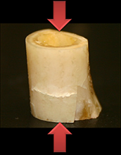
Materials scientists at New York University (NYU) use a compression machine to study how bones split and shatter. The results may ultimately help physicians better diagnose and treat injuries and aid engineers as they improve protection for military and civilian armor, including helmets. This image depicts the fracture of a rabbit femur bone at a slow (quasi-static) compression rate. The bone is showing cracks due to compression.
Credit: Vasanth C. Shunmugasamay, NYU-Poly
Download the high-resolution JPG version of the image. (165 KB)
Use your mouse to right-click (Mac users may need to Ctrl-click) the link above and choose the option that will save the file or target to your computer.
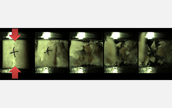
This image shows the fracture of a bone specimen when the compression rate is high; the result is significant fragmentation. The total time duration for this deformation sequence is roughly 1-1,000th of a second.
Credit: Vasanth C. Shunmugasamay, NYU-Poly
Download the high-resolution JPG version of the image. (172 KB)
Use your mouse to right-click (Mac users may need to Ctrl-click) the link above and choose the option that will save the file or target to your computer.
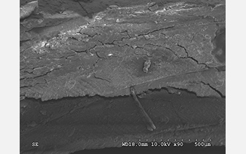
These scanning electron microscope images show significant cracking of a bone specimen that was exposed to high deformation rates. A network of microscopic cracks is apparent.
Credit: Vasanth C. Shunmugasamay, NYU-Poly
Download the high-resolution JPG version of the image. (1.6 MB)
Use your mouse to right-click (Mac users may need to Ctrl-click) the link above and choose the option that will save the file or target to your computer.