
All Images
Research News
2007: Year in Review
In 2007, NSF-supported scientists, engineers and educators announced many notable research and education results and activities. Some of the highlights are presented here.
Credit: Adrian Apodaca, National Science Foundation

The news and Discovery stories that were selected most often by the growing number of visitors to the NSF Web site are also listed.
Credit: Zettl Research Group, Lawrence Berkeley National Laboratory and University of California at Berkeley
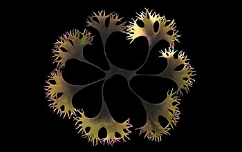
This feathery, dendritic image of Irish moss (Chondrus crispus)--created by Andrea Ottesen, a botanist and molecular ecologist at the University of Maryland, College Park--shared a first place prize in the photography category of the 2007 Science and Engineering Visualization Challenge, sponsored by NSF and the journal Science. To view all of the winners, see http://www.nsf.gov/news/special_reports/scivis/index.jsp?id=win2007.
Credit: Andrea Ottesen, University of Maryland
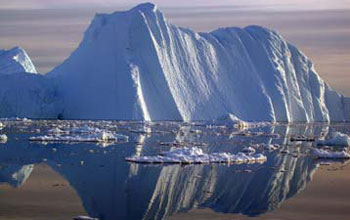
An iceberg calved from a glacier floats in the Jacobshavn fjord in southwest Greenland. A Colorado University-Boulder study indicates Greenland continues to lose ice mass and the rate of loss is accelerating.
Credit: Konrad Steffen, CIRES/University of Colorado at Boulder
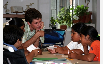
An analysis of K-12 math and science data from schools participating in NSF's Math and Science Partnership program shows steady increases in proficiency.
Credit: Mark Whitmore
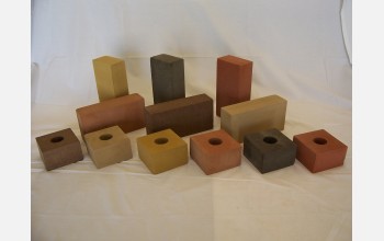
Researchers have developed bricks that look and perform like normal bricks, yet are crafted from fly ash, a waste produced by coal-fired power plants.
Credit: Henry Liu, FPC
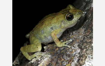
The ancestors of this frog--Eleutherodactylus eunaster--rafted on a voyage across the Caribbean. This frog sits on a tree in Haiti.
Credit: Eladio Fernandez
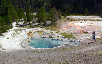
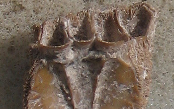
Amaya M. Garcia Costas, a graduate student at Penn State University and a member of the research team that discovered a new chlorophyll-producing bacterium, stands next to colorful microbial mats in Octopus Spring in Yellowstone National Park.
Credit: David Strong, Penn State University
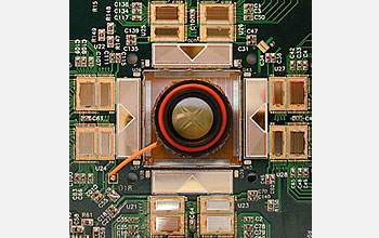
A pinhead size 512-electrode array in the center of this "neuroboard" records the output of retinal neurons.
Credit: Rachel Kalmar and Alan Litke
How do scientists know a whale was eaten here? Whale bones typically are too large for people to carry long distances to archaeological sites. Early humans carried only the skin and blubber. Interestingly, it turns out there are barnacle species that only live on the skin of whales. When people scavenged a beached whale and ate it, all that remained was the barnacle as a sign that says, "a whale was eaten here 164,000 years ago!"
Credit: South African Coast Paleoclimate, Paleoenvironment, Paleoecology, Paleoanthropology Project (SACP4); Arizona State University, Director Curtis W. Marean
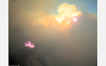
A shot from the real-time, HPWREN-connected camera atop Lyons peak that was taken during the height of the Harris fire, which burned almost 100,000 acres in San Diego County in October 2007.
Credit: HPWREN, funded by the National Science Foundation.
This image, taken by a transmission electron microscope, shows a single carbon nanotube protruding from an electrode. This nanotube is less than a micron long and only 10 nanometers wide, or 10,000 times thinner than the width of a single human hair. When a radio wave of a specific frequency impinges on the nanotube, it begins to vibrate vigorously. An electric field applied to the nanotube forces electrons to be emitted from its tip. This electrical current may be used to detect the mechanical vibrations of the nanotube, and thus listen to the radio waves. (The waves shown in this image were added for visual effect, and are not part of the original microscope image.)
Credit: Zettl Research Group, Lawrence Berkeley National Laboratory and University of California at Berkeley