
Multimedia Gallery
New look at cell membrane
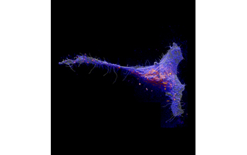
Using a completely new approach to imaging cell membranes, researchers found that a class of molecules called sphingolipids congregate in large patches in the cell membrane. Red and yellow colors indicate local elevations in the sphingolipids abundance.
The study, which revealed some surprising relationships among molecules within cell membranes, was conducted by researchers at the University of Illinois (U of I), Lawrence Livermore National Laboratory and the National Institutes of Health. The research was supported in part by the National Science Foundation (grant CHE 10-58809).
To learn more about this research, see the U of I news story New look at cell membrane reveals surprising organization. (Date of Image: December 2011)
Credit: Courtesy Kevin J. Carpenter
Images and other media in the National Science Foundation Multimedia Gallery are available for use in print and electronic material by NSF employees, members of the media, university staff, teachers and the general public. All media in the gallery are intended for personal, educational and nonprofit/non-commercial use only.
Images credited to the National Science Foundation, a federal agency, are in the public domain. The images were created by employees of the United States Government as part of their official duties or prepared by contractors as "works for hire" for NSF. You may freely use NSF-credited images and, at your discretion, credit NSF with a "Courtesy: National Science Foundation" notation.
Additional information about general usage can be found in Conditions.
Also Available:
Download the high-resolution TIF version of the image. (19.7 MB)
Use your mouse to right-click (Mac users may need to Ctrl-click) the link above and choose the option that will save the file or target to your computer.