
Multimedia Gallery
 All images in this series
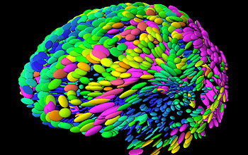
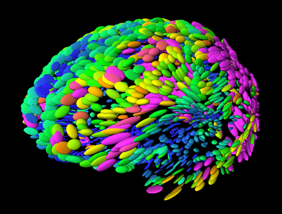
Template brain used to compare with target brains from subjects
This template brain is used in studies on the anatomical differences in brains related to aging and disease. The variation in color and shape of the spheres at left indicates the magnitude and principal directions of the anatomical differences across subjects. In this model, the greatest difference is in the parietal lobe (lower right), an anticipated result since this lobe appeared much later in the evolutionary process than did more primitive brain areas that vary little from brain to brain.
[Research supported in part by U.S. National Science Foundation grant DBI 9601356.]
Credit: Paul Thompson and Arthur Toga, UCLA
Images and other media in the National Science Foundation Multimedia Gallery are available for use in print and electronic material by NSF employees, members of the media, university staff, teachers and the general public. All media in the gallery are intended for personal, educational and nonprofit/non-commercial use only.
Images credited to the National Science Foundation, a federal agency, are in the public domain. The images were created by employees of the United States Government as part of their official duties or prepared by contractors as "works for hire" for NSF. You may freely use NSF-credited images and, at your discretion, credit NSF with a "Courtesy: National Science Foundation" notation.
Additional information about general usage can be found in Conditions.
Also Available:
Download the high-resolution JPG version of the image. (268 KB)
Use your mouse to right-click (Mac users may need to Ctrl-click) the link above and choose the option that will save the file or target to your computer.